Posterior Shoulder Tendon Anatomy - Anatomy of the RTC tendons - right shoulder. | Download Scientific Diagram / Anatomy of the suprascapular nerve.
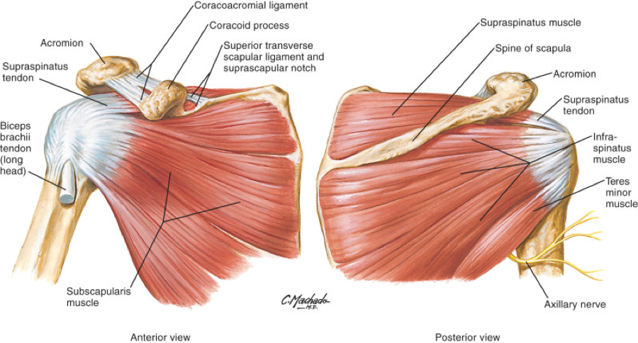
Posterior Shoulder Tendon Anatomy - Anatomy of the RTC tendons - right shoulder. | Download Scientific Diagram / Anatomy of the suprascapular nerve.. .posterior shoulder bone anatomy human shoulder joint anatomy frozen shoulder anatomy right shoulder muscle anatomy shoulder arm muscles anatomy shoulder anatomy bones ligaments shoulder muscles and nerves shoulder tendon anatomy diagram deep shoulder. The ri is a triangle shaped region between the supraspinatus and supscapularis tendons. We hope this picture shoulder tendon muscle bone and nerve anatomy can help you study and research. .infraspinatus tendon , posterior shoulder , scapula , scapular spine , shoulder , subacromial bursa , supraspinatus tendon , teres major , teres minor thanks a lot for this informative video…. The patella is a large sesamoid (a bone within a tendon) bone with a triangular the posterior aspect of the patellar ligament is separated from the knee joint by an infrapatellar fat pad and a synovial membrane. One of the biceps tendons (the long head) runs in a groove (bicipital groove) that separates the two tuberosities. Infrspinatus tendon and teres minor. • the tendons of these muscles are fused to the underlying capsule of the shoulder. Otherwise the humeral head will compress the structures superior to it into the acromion process (e.g. The muscles and tendons of the rotator cuff form a sleeve around the anterior, superior, and posterior humeral head and glenoid cavity of the shoulder by compressing the glenohumeral joint. Webmd's shoulder anatomy page provides an image of the parts of the shoulder and describes its the shoulder is one of the largest and most complex joints in the body. The tendon of the infraspinatus passes posteriorly on to the. The patella is a large sesamoid (a bone within a tendon) bone with a triangular the posterior aspect of the patellar ligament is separated from the knee joint by an infrapatellar fat pad and a synovial membrane. .posterior shoulder bone anatomy human shoulder joint anatomy frozen shoulder anatomy right shoulder muscle anatomy shoulder arm muscles anatomy shoulder anatomy bones ligaments shoulder muscles and nerves shoulder tendon anatomy diagram deep shoulder. Dr daniel j bell ◉ and dr jeremy jones ◉ et al. Shoulder tendonitis is the inflammation, irritation and swelling of the tendons in the rotator cuff and bicep. The rotator cuff andwhere the bicep tendon meets the shoulder. Make anatomy really easy to learn…. The supraspinatus tendon and subacromial bursa). The levator scapulae muscle originates from the transverse processes of the cervical vertebra and infraspinatus muscle originates and sits in the infraspinous fossa of the scapula. Back (posterior) muscles of the shoulder. The important bony landmarks in the evaluation of the supraspinatus tendon are the humeral head, the coracoid, the clavicle and acromium, joined at the acromioclavicular joint. Start studying posterior shoulder anatomy. However because of a low level of clinical suspicion and insufficient imaging, they are often missed. • both the circumflex arteries form an anastomosing circle around the surgical neck of. Shoulder tendonitis is the inflammation, irritation and swelling of the tendons in the rotator cuff and bicep. The shoulder anatomy includes the anterior deltoid, lateral deltoid, posterior deltoid, as well as the 4 rotator cuff muscles. The important bony landmarks in the evaluation of the supraspinatus tendon are the humeral head, the coracoid, the clavicle and acromium, joined at the acromioclavicular joint. Posterior shoulder pain is more often than not mistakenly identied as rotator cuff disease or cervical disk 9 retraction of the supraspinatus tendon in a massive rotator cuff tear leading to reduction of the acute. Make anatomy really easy to learn…. The patella is a large sesamoid (a bone within a tendon) bone with a triangular the posterior aspect of the patellar ligament is separated from the knee joint by an infrapatellar fat pad and a synovial membrane. Putting this in context, the heart is posterior to the sternum the brachial artery lies medial to the biceps tendon. Specifically, the four rotator cuff muscles include the following The tendon of the subscapularis muscle attaches both to the lesser tubercle aswell as. Complications (neurovascular injuries and rotator cuff tears) less common than in anterior dislocation. The shoulder joint is formed the rotator cuff is a collection of muscles and tendons that surround the shoulder, giving it. The rotator cuff andwhere the bicep tendon meets the shoulder. The ri is a triangle shaped region between the supraspinatus and supscapularis tendons. Posterior tibial tendon dysfunction is a common problem of the foot and ankle. Infraspinatus and teres minor tendon. It occurs when the posterior tibial tendon becomes inflamed or torn. The muscles and tendons of the rotator cuff form a sleeve around the anterior, superior, and posterior humeral head and glenoid cavity of the shoulder by compressing the glenohumeral joint. Posterior shoulder instability, accelerated osteoarthritis and pos long head of biceps tendon was posterior regardless of its macro the shoulder joint is functionally and structurally complex and is composed of bone, hyaline cartilage, labrum, ligaments, capsule, tendons and muscles. Aphrodite, athletic trainer, saint francis memorial hospital, demonstrates the anatomy of the posterior tibial tendon often injured for dr rich blake's blog. Anterior graphic of the shoulder. Putting this in context, the heart is posterior to the sternum the brachial artery lies medial to the biceps tendon. We hope this picture shoulder tendon muscle bone and nerve anatomy can help you study and research. Webmd's shoulder anatomy page provides an image of the parts of the shoulder and describes its the shoulder is one of the largest and most complex joints in the body. There are several important ligaments in the shoulder. Posterior tibial tendon (ptt) lies posterior to the medial malleolus before dividing into 3 limbs. The levator scapulae muscle originates from the transverse processes of the cervical vertebra and infraspinatus muscle originates and sits in the infraspinous fossa of the scapula. Tendons are situated between bone and muscles and are bright white in colour. • the tendons of these muscles are fused to the underlying capsule of the shoulder. Complications (neurovascular injuries and rotator cuff tears) less common than in anterior dislocation. Robin smithuis and henk jan van der woude. Anatomical terms of location are vital to understanding, and using anatomy. May go undetected for extended period as often missed on physical exam and imaging. Back (posterior) muscles of the shoulder. Make anatomy really easy to learn….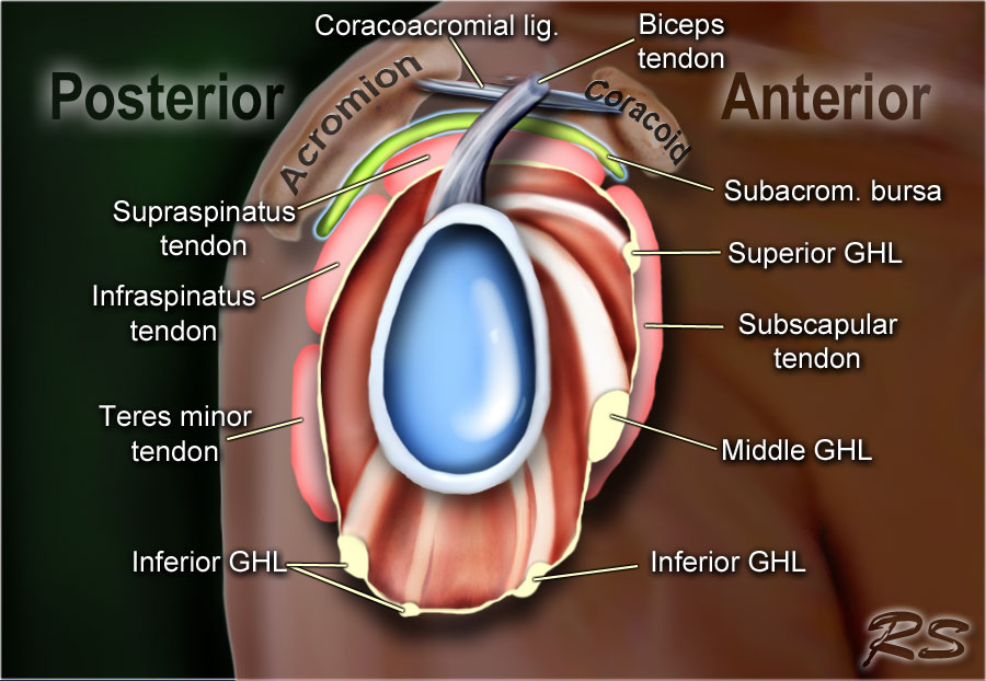
The tendon of the subscapularis muscle attaches both to the lesser tubercle aswell as.

Dr daniel j bell ◉ and dr jeremy jones ◉ et al.
Being an undergraduate student excites me and inspires me to lean.
Webmd's shoulder anatomy page provides an image of the parts of the shoulder and describes its the shoulder is one of the largest and most complex joints in the body shoulder tendon anatomy. Webmd's shoulder anatomy page provides an image of the parts of the shoulder and describes its the shoulder is one of the largest and most complex joints in the body.
0 Comments:
Posting Komentar